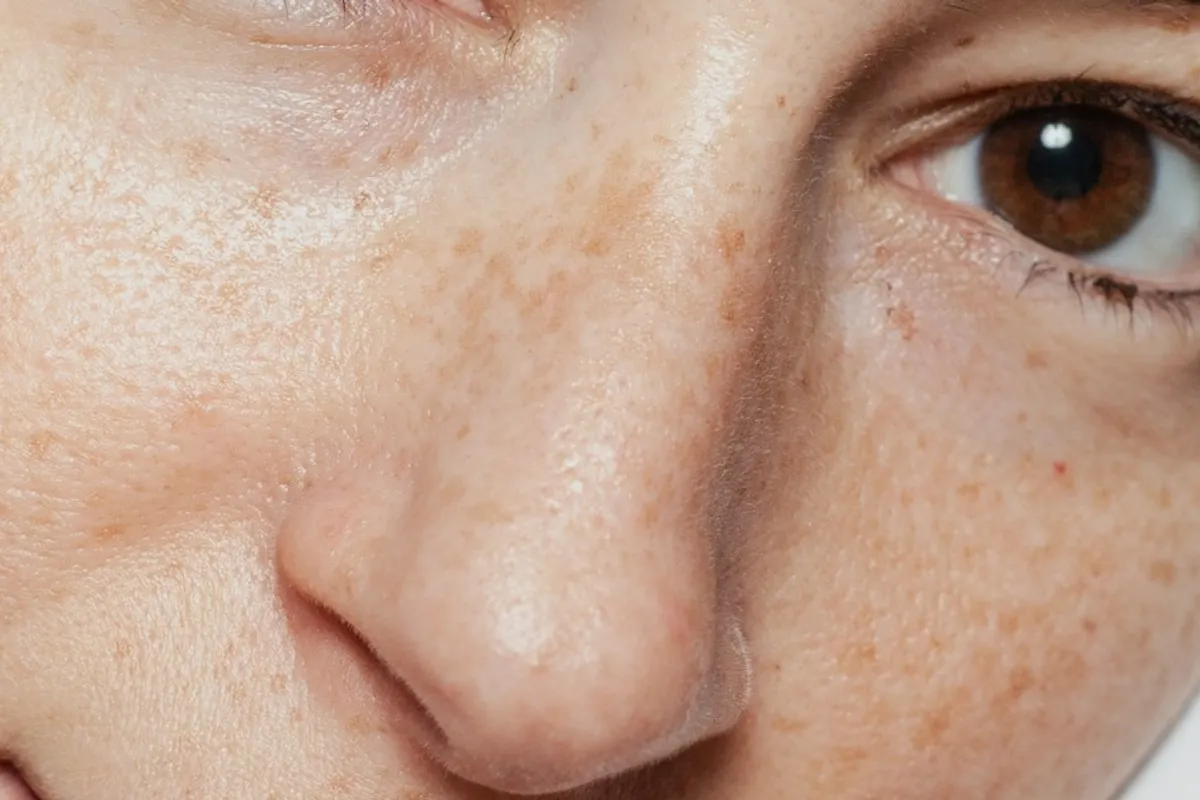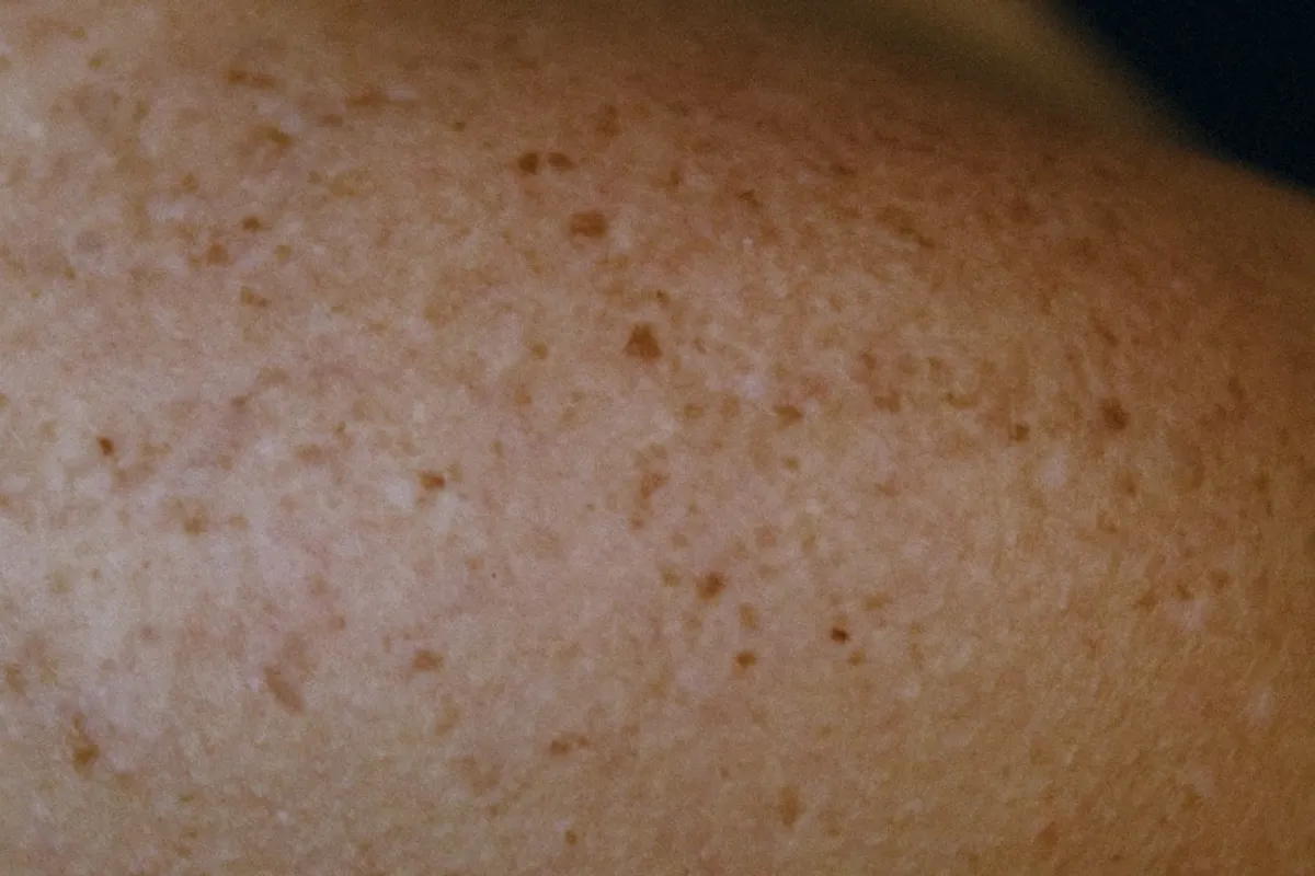
Know what is usual for your skin
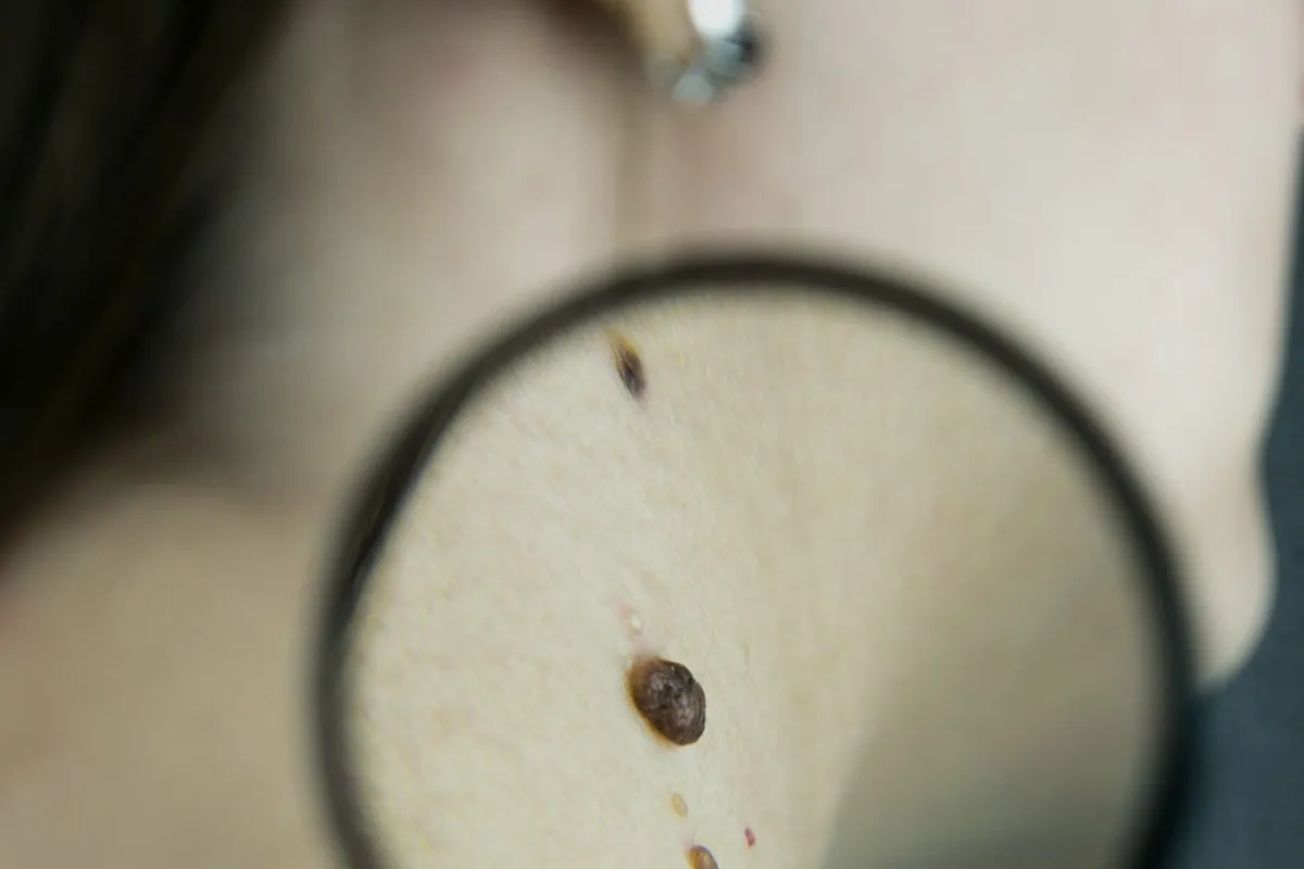
A new spot, a changing mole or a mark that looks different from your others deserves attention. A suspicious change can be painless. Remember areas that are difficult to see, such as your back, scalp and soles; a mirror or someone you trust can help you look.
Use ABCDE as a prompt
Look for asymmetry, an irregular border, different colours, diameter and evolution. Melanomas are often larger than 6 mm, but smaller lesions can also matter. Changes in size, shape, colour or sensation should be assessed. Bleeding without an obvious injury or a sore that does not heal also warrants medical advice.
This checklist is an observation aid, not a way to rule out cancer. You do not need to find every sign before arranging an examination.
What dermoscopy adds
A dermatoscope combines lighting and magnification to reveal structures below the visible surface. The examination is non-invasive and may be performed with or without skin contact. Its findings are considered alongside the history and clinical examination.
The next step may be photographic monitoring, further investigation or removal for histological analysis. Dermoscopy does not provide an absolute guarantee or replace a biopsy when one is needed. Follow-up intervals depend on individual findings and risk.
Prepare for your appointment
Note when the change began, any itching or bleeding and a family history of melanoma. Bring earlier photographs, examination reports and histology results if available. Point out the exact spot that worries you.
Phone photographs taken at similar distances and lighting can help document change, but they cannot replace an examination. Avoid home acids or cosmetic removal of a suspicious lesion before medical assessment.